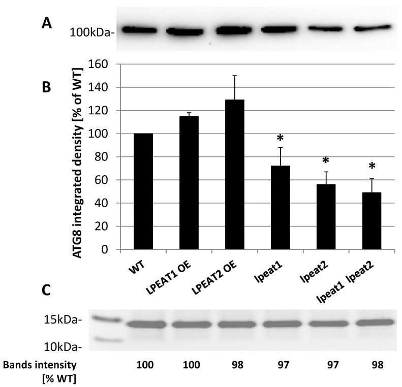
Figure 7.
(A) AGT8-selected western blot analysis of the total protein extracts from leaves (0 DAF) of wild-type Arabidopsis plants (Col-0) and LPEAT1 and LPEAT2 overexpressors, lpeat1 and lpeat2 single mutants and lpeat1 lpeat2 double mutant grown in soil under standard conditions. The blot was incubated with polyclonal antibodies (anti-APG8a/ATG8a (abcam)) against AtATG8 protein. Equal amounts of protein (10 µg) were loaded and separated on NuPAGE 4–12% Bis-Tris-Gel. (B) ATG8 band average intensity (means ± SD) from 4 to 5 independent experiments as a percentage of WT. Asterisk indicates significant difference compared with LPEAT1 OE in “mean difference two sided test” at α = 0.05. (C) Fragment of selected gel with stained protein bands localized between 15 and 10 kDa and “Image Lab 6.01” analyses of the intensity of these bonds.

