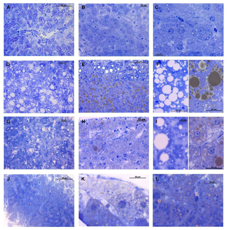
Figure 5.
Toluidine blue staining of liver semithin sections. Control group shows a regular hepatic morphology with few little drops of unsaturated lipids (brown-colored) visible within the hepatocyte’s cytoplasm (A–C). HFD mice show a noticeable hepatosteatosis condition, characterized by hepatocytes with a wide intracytoplasmic spread of both saturated (white-colored) and unsaturated lipid drops of various sizes (D–F). DMG-treated mice fed with HFD show improvement in steatosis, reduction in fat deposits and substantial decrease in lipid drop size (G–I). DMG-treated mice show a slight increase in the size of lipid drops, compared to the control group (J–L). Magnification 40× in (A,B,D,E,G,H,J,K); 100× in (C,F,I,L).

