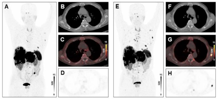
Figure 3.
Absence of treatment response to PRRT in a patient with high systemic inflammation at baseline. MIP image (A) and transversal PET/CT images (B–D) of baseline [68Ga]Ga-DOTA-TATE-PET/CT showing osseous and hepatic metastases in a 76-years-old male GEP-NET patient. MIP image (E) and transversal PET/CT images (F–H) at follow-up after two cycles of PRRT demonstrating progression with new osseous lesions (SSR-TV decreased mildly from 797.26 cm3 to 734.09 cm3, however new lesions appeared, consistent with progressive disease). The baseline CRP level was 31.2 mg/L, the PCM was 14,196, and the HSPI 1, evidencing high systemic inflammation.

