Figure 3.
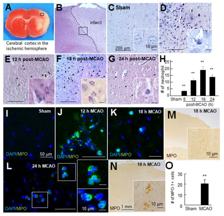
Neutrophils were infiltrated in the post-MCAO brains. (A–H) Coronal brain sections were prepared at 6, 12, 18, or 24 h post-MCAO and stained with Hematoxylin and Eosin. Numbers of neutrophils were counted in the cerebral cortex indicated by the black box (0.5 × 0.5 mm2). Representative pictures are shown (B–G), and results are presented as mean ± SEM (n = 12 from 3 animals) (H). (I–L) Coronal brain sections were prepared from the sham-operated control group and MCAO group at 12, 18, or 24 h post-MCAO and stained with anti-myeloperoxidase (MPO) antibody and 4′,6-diamidino-2-phenylindole (DAPI). Representative pictures of the cortex are shown. (M–O) Coronal brain sections were prepared from the sham control group and MCAO group at 18 h post-MCAO and stained with anti-MPO antibody, and numbers of MPO-positive cells were counted in the indicated black box (0.5 × 0.5 mm2). Scale bars in (C–G) and (I–L) represent 200 and 50 μm, respectively, and those in insets represent 10 μm. Sham, sham-operated rats; MCAO, PBS-treated MCAO controls. ** p < 0.01 vs. sham control by one-way ANOVA with Student–Newman–Keuls test.

