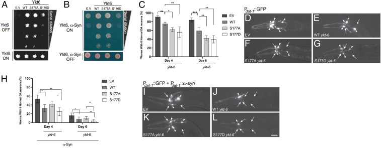
Fig. 6.
Regulation of the evolutionarily conserved CaN-dependent site is critical for cellular physiology in both yeast and C. elegans models of α-synuclein toxicity. (A) WT yeast cells were spotted onto plates containing Synthetic Defined Leu media and episomal Ykt6-Leu selective and then replica plated in fourfold serial dilutions onto episomal Ykt6-inducing plates (Galactose [SGal]-Leu; episomal-Leu selective: empty vector [EV], WT, phosphoablative [S176A], and/or phosphomimetic [S176D]). Endogenous Ykt6 is depleted by incubating the cells at 37 °C, the nonpermissive temperature. Representative plate of n = 3. (B) Yeast strains were spotted onto plates containing uninducing media (Synthetic Defined Leu; Gal-Ykt6, Gal α-syn selective; Lower) and replica platted in fourfold serial dilutions onto Ykt6, α-syn–inducing plates containing selective media and SGal-Leu (Upper). EV is used as control plasmid. Representative plate of n = 3. (C) The extent of DA neurodegeneration in C. elegans strains overexpressing both GFP and ykt-6 variants (WT, S177A, and S177D) in DA neurons under the dat-1 promoter, determined at both days 4 and 6 posthatching. Control worms overexpressed both GFP and the EV backbone containing the dat-1 promoter, with ykt-6 variants omitted. Data are shown as the average of three independent stable transgenic lines for each construct, with each line in triplicate. Error bars indicate SD. One-way ANOVA with a Tukey’s post hoc test. *P < 0.05, **P < 0.01, ***P < 0.001. (D–G) Representative images of C. elegans neurons taken at day 6 posthatching in the backgrounds described in D. The anterior (head) portion of worms are shown, allowing the visualization of the six DA neurons in this region via GFP fluorescence. Arrowheads indicate intact DA neurons. Arrows indicate degenerated DA neurons in a strain overexpressing control vector and GFP (D), WT ykt-6 and GFP (E), phosphoablative S177A ykt-6 and GFP (F), or phosphomimetic S177D ykt-6 and GFP (G). (H) same as in C but in C. elegans strains overexpressing GFP, α-syn, and ykt-6 variants (WT, S177A, and S177D) in DA neurons under the dat-1 promoter, determined at both days 4 and 6 posthatching. (I–L) same as in D–G in a background overexpressing GFP, α-syn, and ykt-6 variants (WT, S177A, and S177D) in a strain overexpressing EV control vector, GFP, and α-syn in the DA neurons where three neurons are missing (I); WT ykt-6, GFP, and α-syn where five DA neurons are missing (J); phosphoablative S177A ykt-6, GFP, and α-syn in the DA neurons where three neurons are missing (K); or phosphomimetic S177D ykt-6, GFP, and α-syn in the DA neurons where five dopaminergic neurons are missing (L).

