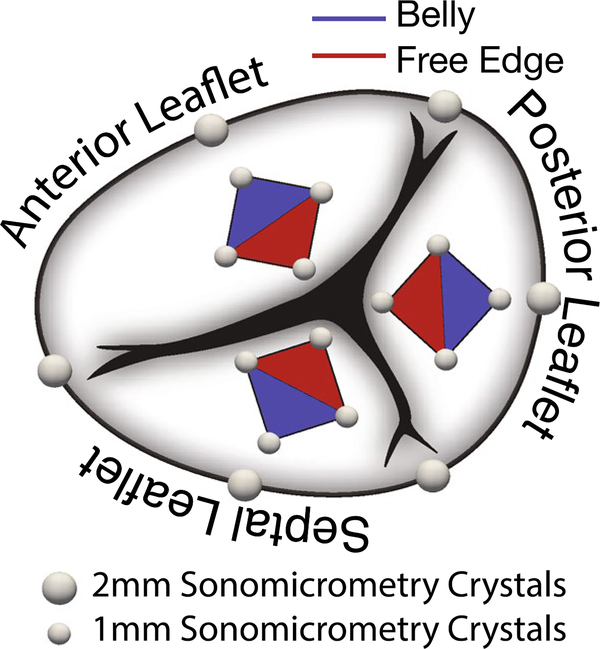
Fig. 1.
Anatomy of the tricuspid valve with sonomicrometry crystal placement. We implanted six, 2-mm crystals along the annulus with three crystals at the commissures and the other three crystals bisecting the resulting annular regions. To determine strains in the leaflet belly (blue) and the leaflet free edge (red), we implanted additional four, 1-mm crystals in a diamond shape on each leaflet for a total of 12 leaflet crystals and 18 valvular crystals

