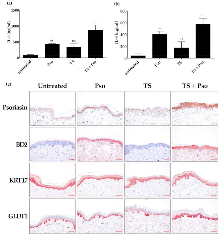
Figure 3.
Generation of an ex vivo psoriasis model. Skin models have been cultured for 4 days at the air-liquid interface using appropriate differentiation medium. The skin models were left untreated, stimulated with psoriasis cytokines (IL-17A, TNF-α, IL-22, 20 ng/mL each; Pso), tape stripped (TS) or both actions were performed (TS + Pso). The protein level of secreted IL-6 (a) and IL-8 (b) was measured in the cell culture supernatant by ELISA (n = 4). (c) Skin models were fixed, embedded in paraffin and 3 µm sections were stained with antibodies against psoriasin (S100A7), β-defensin 2 (BD2), keratin 17 (KRT17) or glucose transporter 1 (GLUT 1) (n = 4). Scale bar = 100 µm. ** p ≤ 0.01, *** p ≤ 0.001, ns-not significant (p > 0.1).

