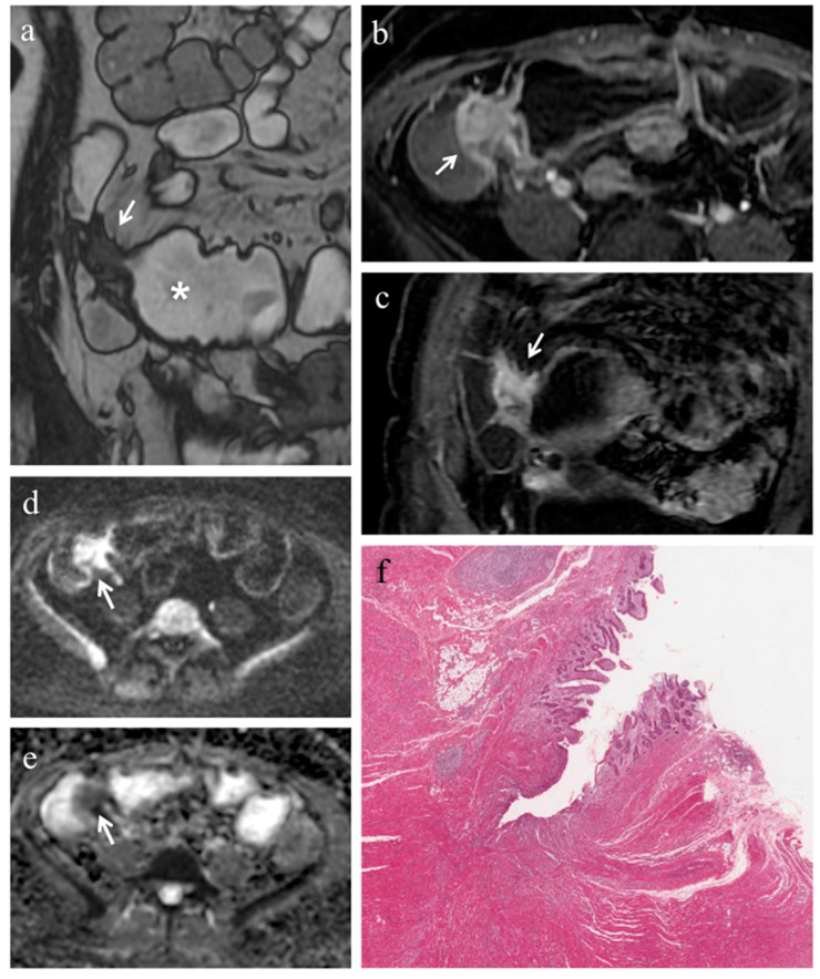
Figure 1.
(a–f). MRE in a 49-year-old woman with CD: severe predominantly fibrotic stricture of the terminal ileum. (a) Coronal fast imaging employing steady state acquisition (FIESTA) image shows wall thickening with noticeable narrowing of the lumen in the terminal ileum (white arrow); note the dilatation (>30 mm) of the upstream bowel loop (white asterisk). (b) Axial and (c) coronal contrast-enhanced fat-suppressed T1-weighted images demonstrate homogeneous enhancement of terminal ileum (white arrows). (d) Axial DW image (b = 800 s/mm2) and (e) corresponding ADC map show the same intestinal segment demonstrating restricted diffusion with high signal intensity (white arrow) on the DWI image and low signal intensity (white arrow) on the ADC map (mean ADC value 0.745 × 10⁻³ mm2/s). (f) Histopathological section from the ileal stricture: hematoxylin and eosin-stained sample (H&E 10×). CD exhibiting severe fibrosis (FS = 2) and moderate inflammation (AIS = 7): muscular layers dissected by dense fibrotic tissue on the left, mucosal ulceration and moderate inflammatory infiltration on the right.

