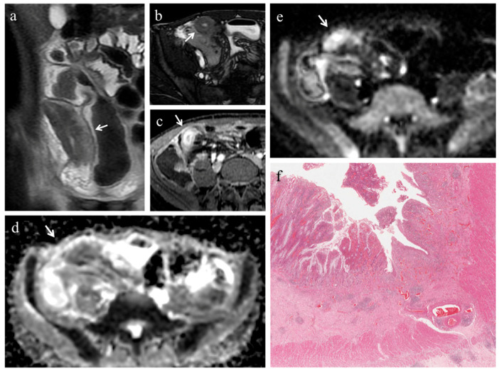
Figure 2.
(a–f). MRE in a 19-year-old man with CD: stenosis of the terminal ileum with concomitant inflammation and fibrosis. (a) Coronal T2-weighted image and (b) axial FIESTA image show marked wall thickening of the terminal ileum with luminal narrowing (white arrows). On (c) axial contrast-enhanced fat-suppressed T1-weighted image, the mural thickening of the terminal ileum displays intense mucosal enhancement (white arrow). The same intestinal segment demonstrates high signal intensity on (d) the DW image (b = 800 s/mm2) and low signal intensity on (e) the corresponding ADC map (white arrows) (mean ADC value 1.096 × 10⁻³ mm2/s), a finding consistent with restricted diffusion. (f) Histopathological section from the ileal stricture: hematoxylin and eosin-stained sample (H&E 10×). CD exhibiting mild/moderate fibrosis (FS = 1) and severe inflammation (AIS = 10): mucosal ulceration and severe inflammatory infiltration on the top; mild/moderate fibrosis, edema and inflammatory infiltration in submucosal layer on the bottom.

