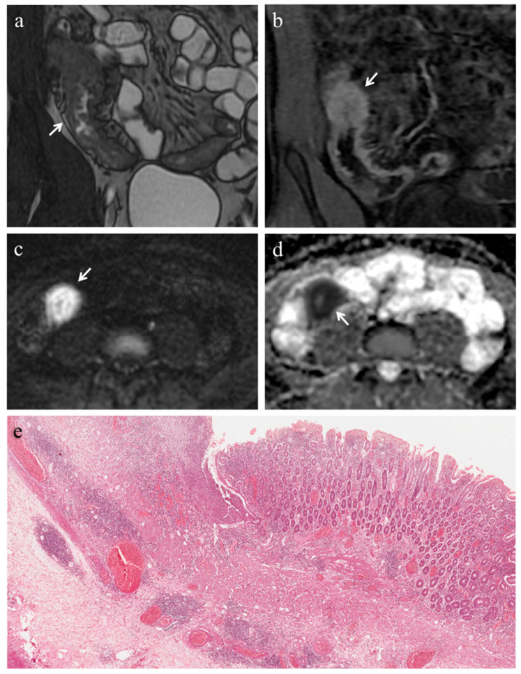
Figure 3.
(a–e). MRE in a 22-year-old man with CD: predominantly inflammatory stricture of the terminal ileum. (a) Coronal FIESTA image shows mural thickening of terminal ileum with stenosis of the lumen (white arrow). (b) Coronal contrast-enhanced fat-suppressed T1-weighted image demonstrates the homogeneous wall enhancement of the affected ileal loop. The same intestinal segment demonstrates restricted diffusion in the form of high signal intensity on (c) the DW image (b = 800 s/mm2) and low signal intensity on (d) the corresponding ADC map (white arrows) (mean ADC value 1.320 × 10⁻³ mm2/s). (e) Histopathological section from the ileal stricture: hematoxylin and eosin-stained sample (H&E 10×). CD exhibiting absent or minimal fibrosis (FS = 0) and severe inflammation (AIS = 9): mucosal ulceration and intense inflammatory infiltration on the top; edema and intense inflammatory infiltration in the submucosal layer.

