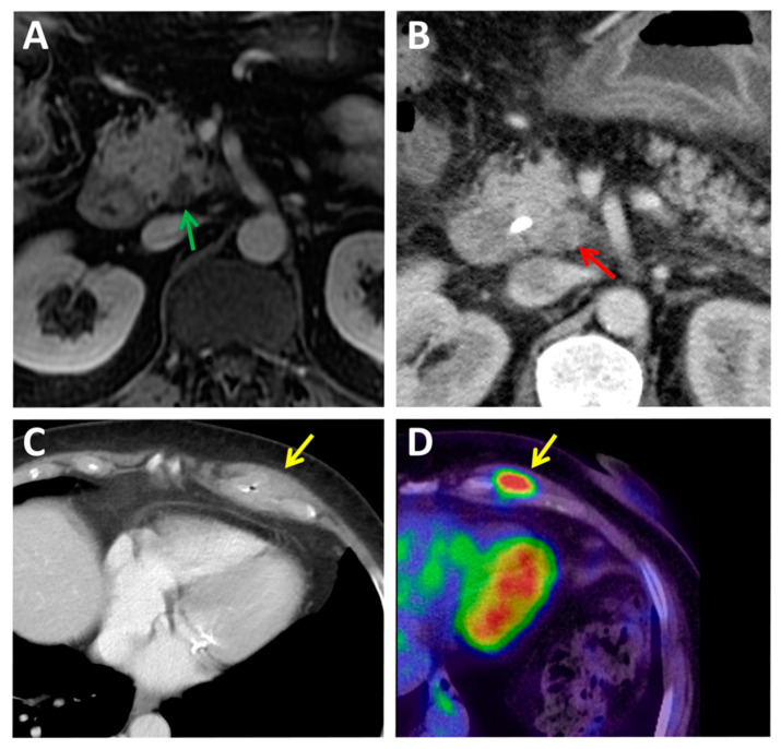
Figure 1.
Cases of radiologically infra-staged and false-positive diagnosis of metastatic tumor recurrence by current imaging methods. (A,B) A radiologically infra-staged T2 N0 pancreatic ductal adenocarcinoma (PDAC) was found to be a locally advanced pT4 pN1 PDAC in the pathology report. (A) magnetic resonance imaging (MRI) showing a small cystic area in the pancreas’ uncinate process (green arrow), a bile duct stricture, no direct signs of malignancy. (B) A computed tomography (CT) performed after bile stent placement showed a small hypodensity (red arrow) next to the superior mesenteric artery. (C,D) A case of a false-positive diagnosis of metastatic tumor recurrence. (C) Some months after surgical excision of a PDAC stage pT2 pN0 M0 R0, an asymptomatic solid mass in the left costal wall (yellow arrow) was shown by a CT scan. (D) An 10.43 SUVmax in 2-[18F]fluoro-2-deoxy-D-glucose ([18F]FDG) positron emission tomography (PET)-CT was suspicious of a PDAC metastatic relapse. A tumor core biopsy found inflammatory and fibrotic tissue but no sign of malignant cells.

