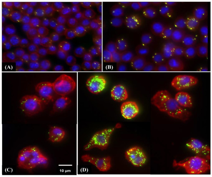
Figure 4.
Immunofluorescence staining of RAW 264.7 macrophages stimulated by HW-Pm. Macrophages were incubated with HW-Pm (100 and 500 µg/mL) and LPS (100 ng/mL) and IFN-γ (5 ng/mL), respectively. Carboxylate-modified polystyrene yellow–green fluorescent beads were added to the cells. Macrophages were fixed with 4% paraformaldehyde, permeabilized using 0.5% Triton X-100 and further stained with Texas Red®-X phalloidin (InvitrogenTM, Waltham, MA, USA) to stain F-actin, and cell nuclei were visualized with 4′,6-diamidino-2-phenylindole (DAPI) under a fluorescence microscope. Representative images of each group (Scale bar 10 µm): (A) Control (DMEM), (B) LPS–IFN-γ, (C) HW-Pm 100 µg/mL and (D) HW-Pm 500 µg/mL.

