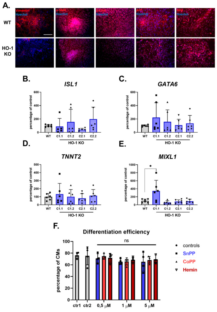
Figure 3.
HO-1 does not influence the efficiency of hiPSCs.2 differentiation to cardiomyocytes. (A) Immunofluorescence analysis of markers of three germ-layers (Vimentin, α-SMA, GATA4, AFP, and NFH) in spontaneously differentiated WT (upper panel) and HO-1 KO (bottom panel) hiPSC.3 via embryoid bodies. Bar indicates 100 µm. qRT-PCR analysis of expression of cardiac mesoderm markers: (B) ISL1, (C) GATA6, (D) TNNT2 and (E) MIXL1. Expression was normalized to EEF-2 levels. (F) Flow cytometric analysis of direct cardiomyocyte differentiation efficiency (based on TNNT2 expression) of WT hiPSC.2 treated with tin protoporphyrin IX (SnPP), cobalt protoporphyrin IX (CoPP) and hemin. Ctr1-DMSO control for SnPP and CoPP, ctr2-2 µM NaOH control for hemin. Bars represent mean ± SD of N = 3 experiments. Dots, squares and triangles represent each replicate for corresponding groups. * p < 0.05, one-way ANOVA test.

