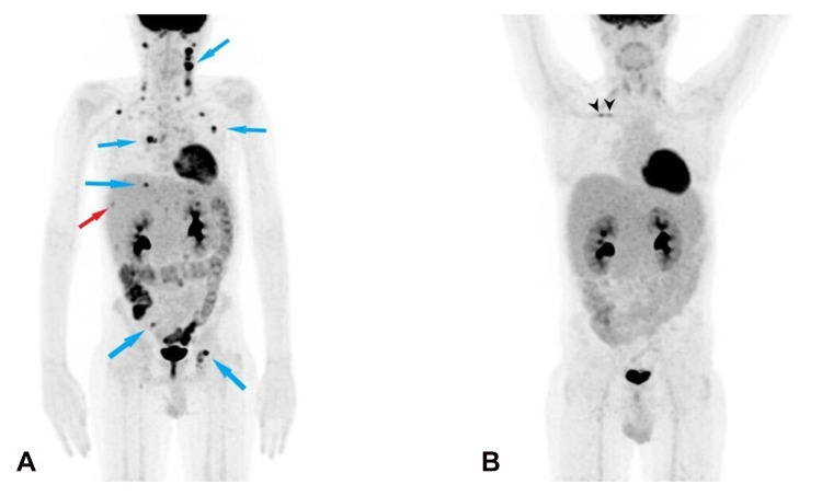
Figure 2.
Baseline (at diagnosis) 18F-FDG PET/CT (A) and post-treatment 18F-FDG PET/CT (B). (A) 18F-FDG PET/CT shows pathological uptake in the following lymph nodes (blue arrows): bilateral laterocervical (SUVmax 21.3 at left station III), bilateral axillary (SUVmax 9.8 in left axilla), right costophrenic recess (SUVmax 6.6), right pulmonary hilum (SUVmax 11.5), celiac, left paraaortic (SUVmax 4.7), right iliac (SUVmax 5.5), bilateral inguinal (SUVmax 11.8 left node) and a pathological uptake at VIs/VIIs liver segment (SUVmax 3.3, red arrow). (B) Post-treatment 18F-FDG PET/CT shows a complete metabolic response (no pathological uptake); two foci uptake can be seen at central venous catheter in right subclavian vein, suspicious for infection (black arrowheads).

