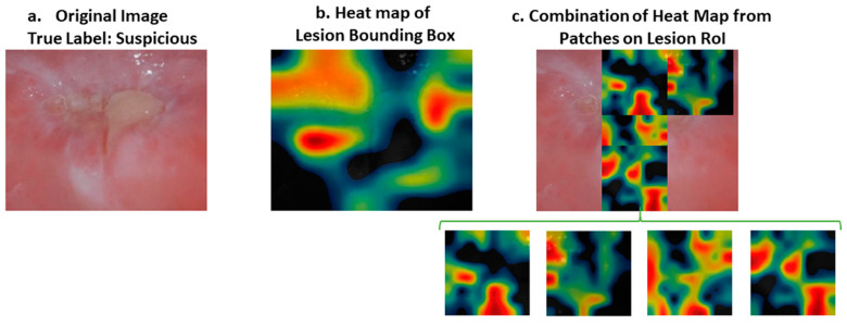
Figure 7.
(a) Original image. (b) Heat map of lesion bounding box. (c) Heat map of a combination of patches belonging to the severe part of the lesion area. Original image was classified as “suspicious” when the region of interest was tested. However, the heat map of the region of interest did not reflect the severe part of the image with a warm color (e.g., red). On the other hand, when we divide the severe part of the image into four patches, the heat map of the patches indicates the severe region with an intense color between yellow and red. RoI: Region of Interest.

