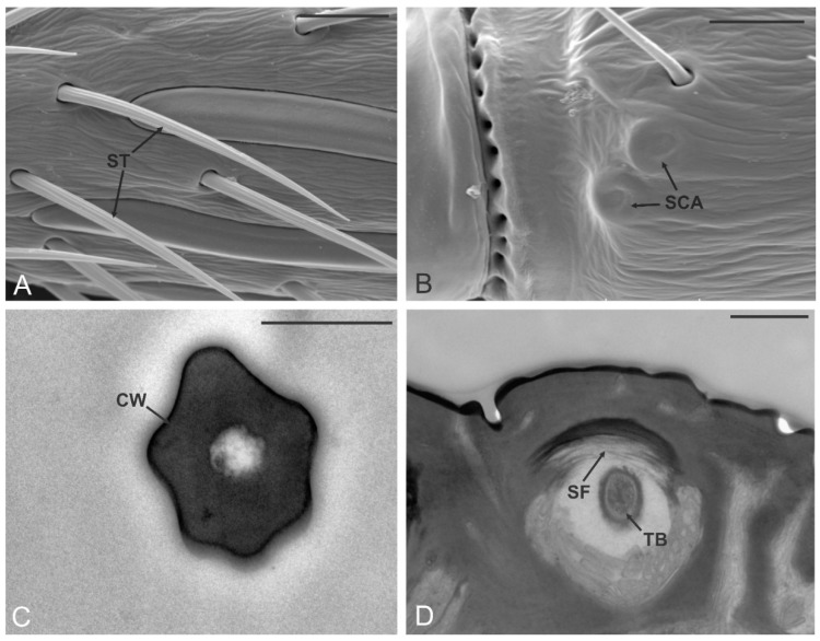
Figure 3.
Sensilla trichoidea (ST) and campaniform sensilla. (A,B) SEM details of the pedicel (A2) and A4 respectively, where it is possible to observe two sensilla trichoidea (ST) and two sensilla campaniformia (SCA). In (C) a TEM cross-section through the cuticular peg of an ST: the thick poreless cuticular wall (CW) delimits an internal lumen without sensory neurons inside. In (D) a TEM section through the socket of ST: suspension fibers (SF) and a single sensory neuron giving rise to a tubular body (TB) are depicted. Bar scale: (A,B), 10 µm; (C), 0.5 µm; (D), 1 µm.

