Figure 4.
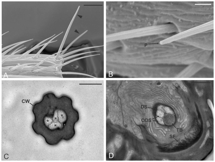
Sensilla chaetica (SCH). (A,B) SEM pictures showing the sensilla chaetica (arrowheads in (A)) typically positioned at the distal margin of one of the antennomeres (in this case A10) and emerging from the rest of the antennal sensilla. In (B) a sensillum chaeticum with the apical pore (P) is highlighted. (C,D) TEM sections of the sensillum revealed the presence of a thick, aporous cuticular wall (CW) and a lumen occupied by four unbranched outer dendritic segments (*). Proximally, at the level of the sensillum socket, sensilla chaetica present suspension fibers (SF) around the socket. The fascicle of sensory neurons is enclosed by a single dendrite sheath (DS) and is made up of the four abovementioned ODS plus a fifth sensory neuron that develops in a tubular body (TB). Bar scale: (A), 10 µm; (B), 2 µm; (C), 0.5 µm; (D), 1 µm.

