Figure 5.
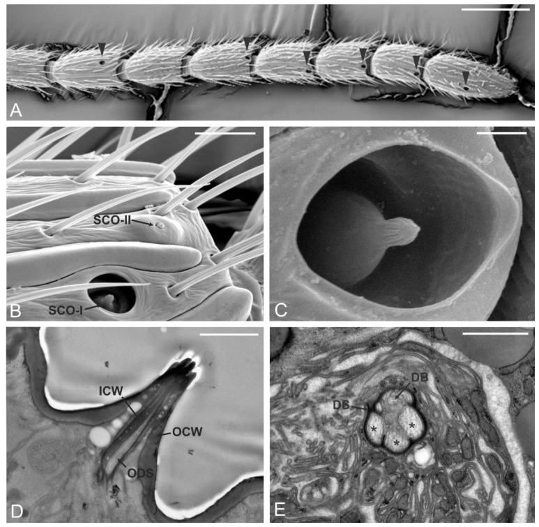
Sensilla coeloconica (SCO). (A) SEM micrographs showing the antennomere interval A8-A14. Each antennomere (except A9) presents a sensillum coeloconicum type I (SCO-I) positioned distally on the antennomere (black arrowheads). (B) Detail of the distal region of A8, SCO-I can be easily distinguished from the sensillum coeloconicum type II (SCO-II), positioned more distally. (C) Close-up view of SCO-I: a grooved peg resides inside the pit that opens externally through a large aperture. (D,E) TEM micrographs of SCO-I. In (D) a longitudinal section through the peg shows the presence of a double wall organization, with an internal cuticular wall (ICW) and an external cuticular wall (OCW). The internal lumen is filled with outer dendritic segments (ODS) of the sensory neurons. In (E) a cross-section taken below the cuticular peg: three ODS (*) and a fourth organized into dendritic branches (DB) are visible, all of them enclosed in a single dendritic sheath (DS). Bar scale: (A), 100 µm; (B), 10 µm; (C,D), 2 µm; (E), 1 µm.

