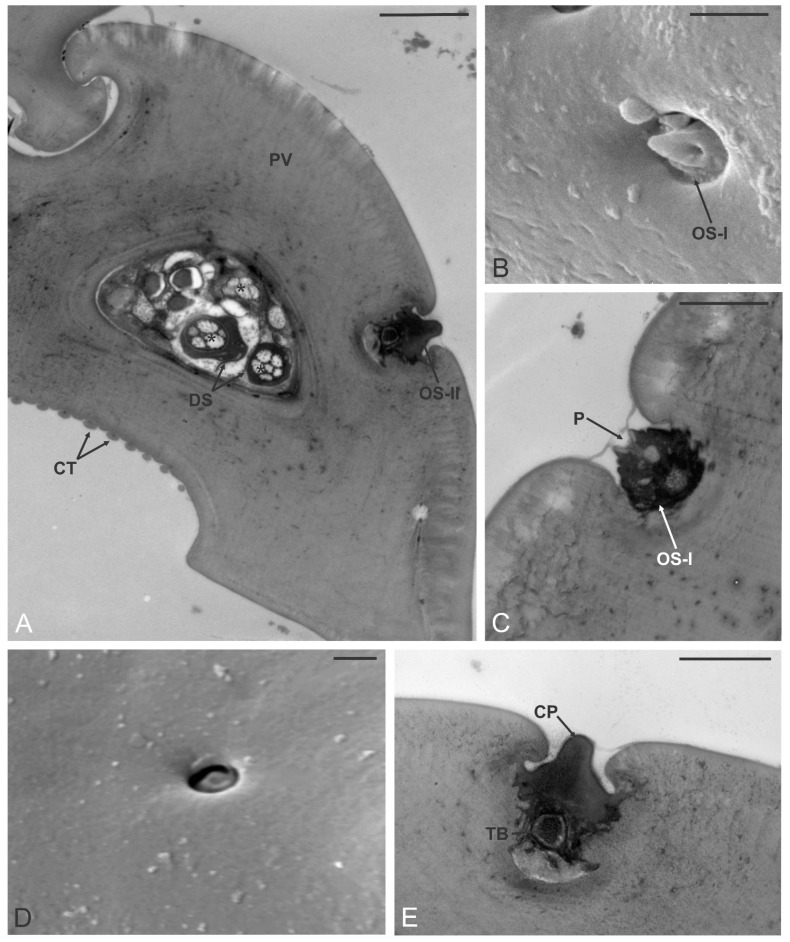
Figure 7.
Dryocosmus kuriphilus ovipositor. (A) TEM cross-section of one of the paired valves (PV): the external side presents a single ovipositor sensillum type II (OS-II), while the internal side is occupied by a single row of ctenidia (CT). The PV lumen reveals the presence of three bundles of sensory neurons (*), each one comprised of 5–6 sensory neurons enclosed in a thick dendrite sheath (DS). (B) SEM close-up view of an OS-I: a well evident apical pore is observed. (C) TEM longitudinal section of OS-I: the apical pore (P) is revealed. (D) SEM picture of an OS-II. (E) TEM longitudinal section of an OS-II: the peg (CP) is made of solid cuticle without pores and reveals the presence of a single sensory neuron ending in a tubular body (TB). Bar scale: (A), 2 µm; (B–E), 1 µm.

