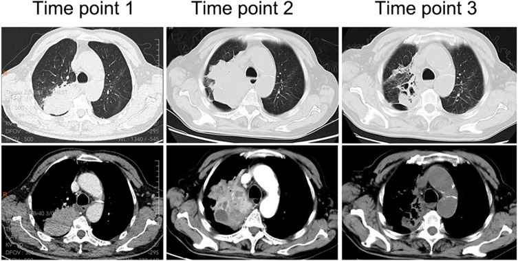
Figure 2.
Lung CT manifestation of case 4. Time point 1: At the first visit, a mass lung consolidation shadow was visible in the right upper lobe. Time point 2: At 8 months of onset, a notably enlarged mass is visible, with liquefaction necrosis in the middle of the lesion. Time point 3: After effective treatment for 3 months, the lesion was significantly smaller with residual cavities and fibrous lesions.

