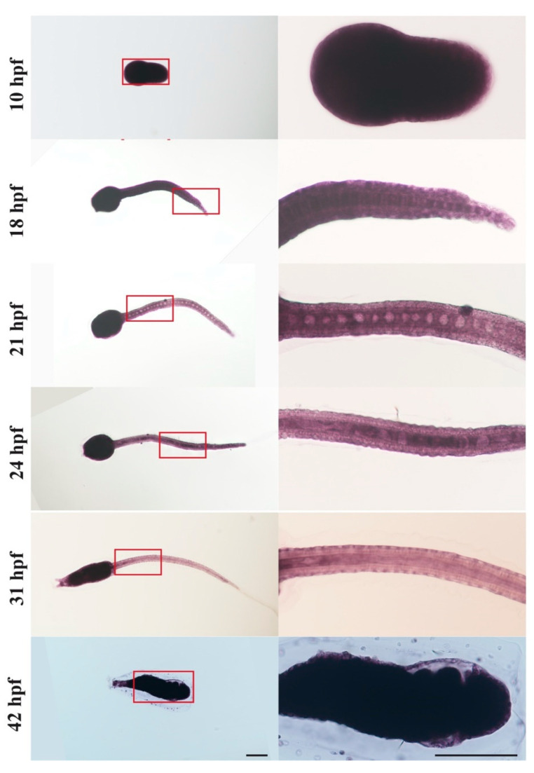
Figure 5.
Expression patterns of csa-miR-92c detected by in situ hybridization. Embryos and larvae at 10, 18, 21, 24, 31, and 42 hpf were hybridized with locked nucleic acid (LNA) probes of csa-miR-92c. The developmental stages were indicated. The signals of csa-miR-92c were detected in the whole body at 10 hpf and 42 hpf and expressed in the trunk, notochord cells, and epithelial cells at 18, 21, 24, and 31 hpf. The red frame indicated the regions of zoom-in images in the middle column. Scale bars represent 100 μm.

