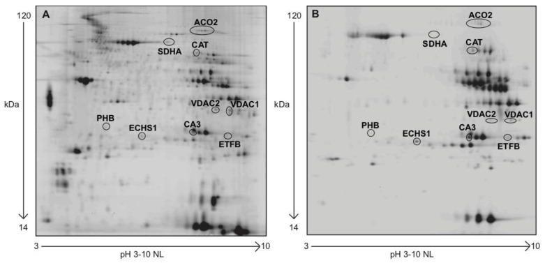
Figure 3.
Representative VL muscle protein nitroprofiles. (A) Individual 2D pattern gel image of 40 μg of protein extract, separated in a pH 3–10 non-linear immobilized pH-gradient (IPG) in the first dimension, and SDS gel (12% T, 2.5% C) in the second dimension. (B) NITRO-DIGE gel image of 20 μg of protein extract, separated in a pH 3–10 non-linear IPG strip in the first dimension and SDS gel (12% T, 2.5% C) in the second dimension.

