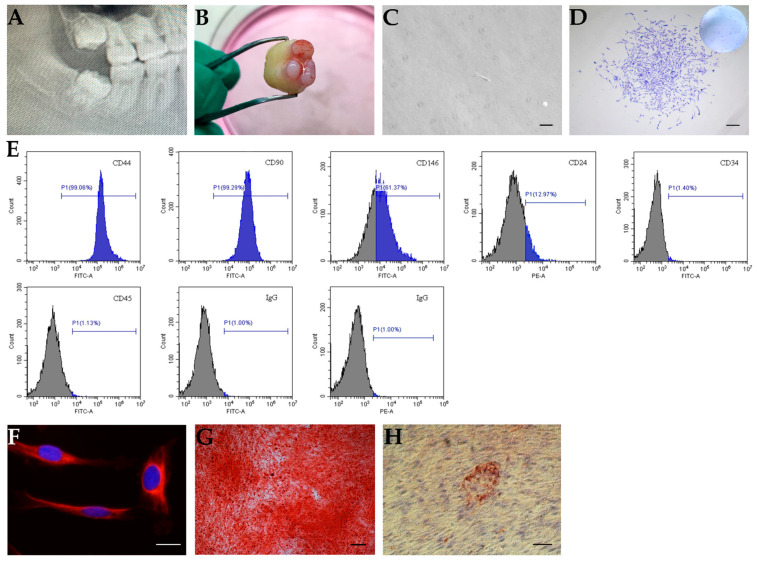
Figure 1.
In vitro characterization of stem cells from apical papilla (SCAPs). (A,B) The apical papilla tissue from an immature human tooth. (A) A representative radiograph indicating the third molar with the open apex; (B) apical papilla tissue. (C,D) The colony formation of SCAPs. (C) SCAPs exhibited a typical spindle shape, scale bar = 100 μm. (D) Isolated cells formed colony-like units, scale bar = 200 μm. (E) Flow cytometric analysis of SCAPs using several markers. Cells were positive for CD44, CD90, CD146, and CD24, and negative for CD34 and CD45. (F) Immunofluorescence staining showed nestin expression in SCAPs, scale bar = 20 μm. (G) SCAPs formed mineralized nodules after osteo-/odontogenic differentiation, scale bar = 200 μm. (H) SCAPs were differentiated into adipocytes with Oil Red O-positive lipid clusters when cultured in adipogenic differentiation medium for four weeks, scale bar = 50 μm.

