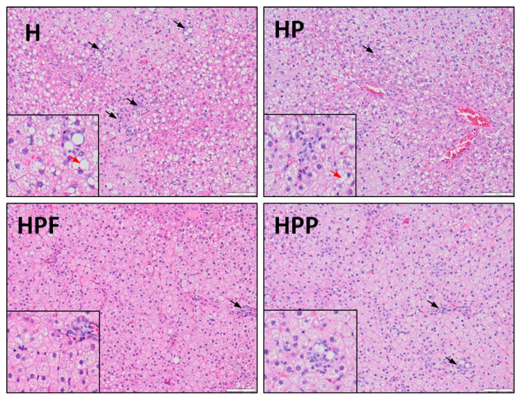
Figure 3.
Hepatic histology in rats fed the experimental diets. H, control high-fat diet; HP, control high-fat diet enriched with raspberry polyphenol extract; HPF, control high-fat diet enriched with raspberry polyphenol extract and fructo-oligosaccharides; HPP, control high-fat diet enriched with raspberry polyphenol extract and pectin. (H) Liver with steatosis. Several inflammatory foci within the hepatic lobule (black arrow) and enhanced hepatocyte ballooning (red arrow). (HP) Reduced amount of inflammatory cells (black arrow) and mild ballooning of cells. Hepatocytes have rounded contours with clear reticular cytoplasm (red arrow). (HPF) There is one area of inflammatory cells (black arrow), the cytoplasm is pink and granular, and liver cells have sharp angles. (HPP) There are several inflammatory foci within the hepatic lobule (black arrow), and the shape of liver cells is quite similar to that observed in the HPF group. The liver samples were stained with haematoxylin and eosin at 20× and 40× (small image) magnification.

