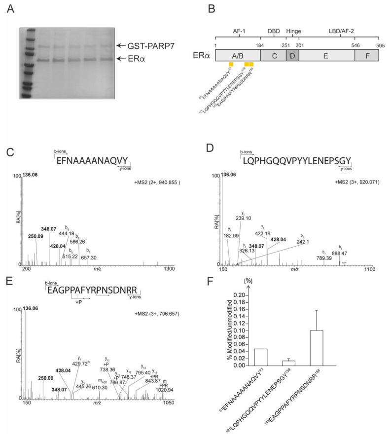
Figure 6.
Identification of mono-ADP-ribosylated peptides in bacterial expressed and purified ERα. (A) Representative SDS-PAGE of GST-PARP7 and ERα prior to LC/MS analysis. (B) A schematic representation of the domain structure of ERα. Location of ADP-ribosylated peptides are denoted by yellow rectangles. Peptide sequences are numbered from the unmodified full-length protein. (C) The MS2 spectrum of the trypsin generated ion at m/z 940.855. (D) The MS2 spectrum of the trypsin generated ion at m/z 920.705. (E) The MS2 spectrum of the trypsin generated ion at m/z 796.657. (F) Relative levels of modification (in percentage) of ADP-ribosylated peptides identified by M/S in ERα.

