Figure 4.
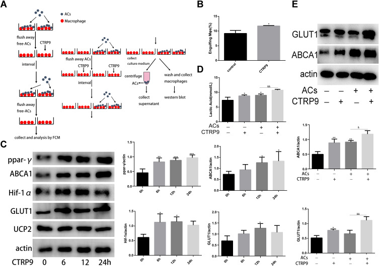
CTRP9 improved macrophage immunometabolism (A) The detailed procedure of two-stage efferocytosis assay. (B) Macrophages were incubated with ACs at a ratio of 10:1 ACs: macrophages for 120 min. The ACs were removed, and then, after a 12h interval with or without CTRP9 treatment, the macrophages were incubated with TAMRA-CypHer5E-labeled ACs at a ratio of 10:1 for 45 min. The unengulfed ACs were removed, and efferocytosis was quantified as the total percentage of macrophages that were positive for TAMRA-CypHer5E-labeled ACs by flow cytometric analysis. (C) Macrophages were stimulated with 1 μg/mL of CTRP9 for 0–24 h, and cell lysates were analyzed for PPAR-y, ABCA1, HIF-1a, GLUT1 and Ucp2 by Western blot. (D and E) Macrophages were incubated with ACs at a ratio of 10:1 ACs: macrophages for 120 min. The ACs were removed, and then, after a 12 h interval with or without CTRP9 treatment. Lactate was measured in the medium. Representative Western blot analyses and quantitative data are shown. All data are expressed as mean ± SD (n=3). *p< 0.05; **p<0.01; ***p<0.001; versus control. &p<0.05; &&p<0.01; versus ACs group.

