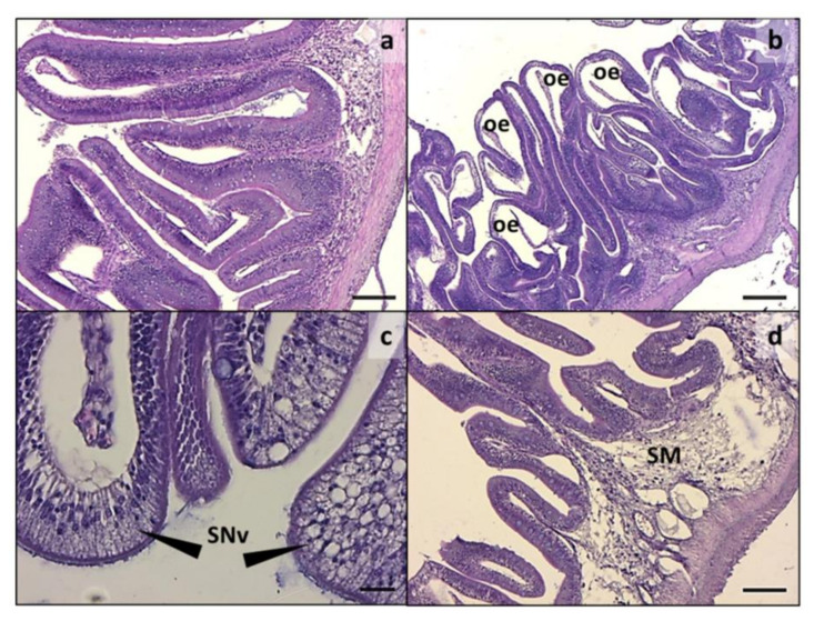
Figure 1.
Medium intestine. (a) Normal histology from CF; (b) histological architecture alteration, including oedema and severe folds fusion degree in CV; (c) high magnification showing enterocyte ipervacuolization in CV; (d) highly infiltrated and vacuolated submucosa in CV group. oe: oedema; SNv: supranuclear vacuoles; SM: submucosa. Scale: a,d = 100 µm; b = 200 µm; c = 20 µm.

