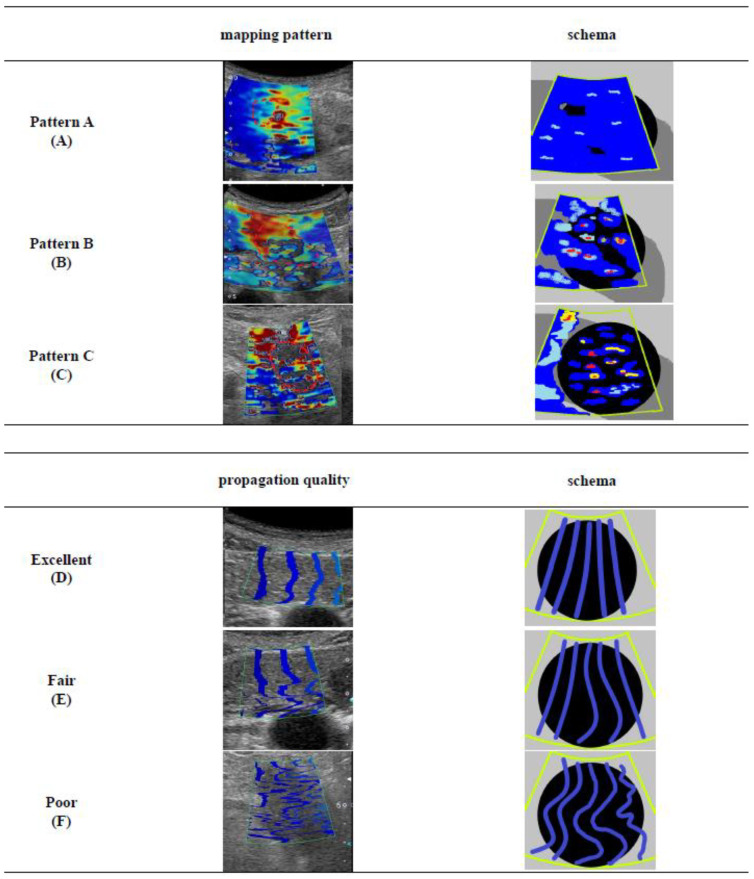
Figure 1.
The definitions of shear wave elastography (SWE) mapping patterns and propagation qualities: (A) Mapping pattern A, whole coloring over the tumor/non-tumor lesion; (B) Mapping pattern B, presence of partial coloring area (major color-displayed area showing 50% or more of the target lesion) and small isolated coloring spots (major color-displayed area showing less than 50% of the target lesion) and uncolored in the other part; (C) Mapping pattern C, multiple small isolated coloring spots (major color-displayed area showing less than 50% of the target lesion) with uncolored areas; (D) Propagation quality “Excellent”, linear contour lines are with equally spaced intervals; (E) Propagation quality “Fair”, some of the lines are showing unequally spaced and/or non-linear appearance; (F) Propagation quality “Poor”, all lines are demonstrating a non-linear appearance with non-linear shaped lines.

