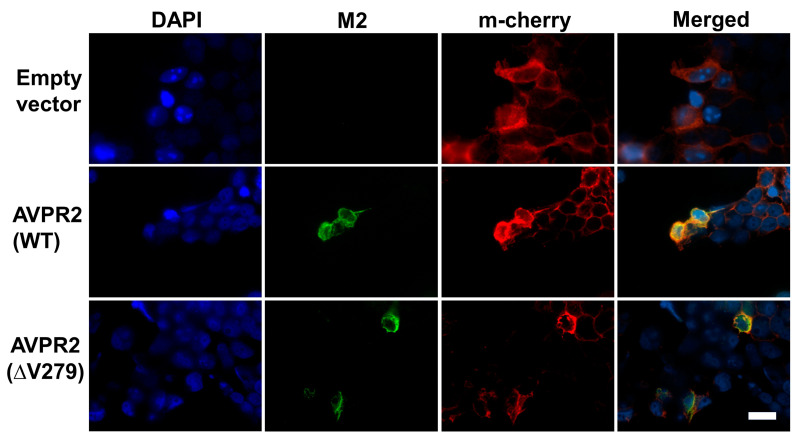
Figure 2.
Arginine vasopressin receptor 2 (AVPR2)-∆V279 is localized in the membrane surface. The Flag-epitope tagged AVPR2 or AVPR2-∆V279 expression vector was co-transfected with a membrane-bound cherry fluorescent protein (m-Cherry) vector into HEK 293T cells. M2 antibody was used for immune-staining, and the locations of AVPR2 proteins were visualized by rhodamine-conjugated secondary antibodies. m-Cherry marked the plasma membrane are shown red, and the AVPR2 proteins are in green. Cell nuclei were counterstained by DAPI (blue). Both single staining and merged images are shown. Both the transfected wild-type and ΔV279 mutant AVPR2 proteins were successfully expressed in HEK 293T cells, and both of the proteins can associate with the plasma membrane as indicated by m-cherry co-localization (yellow) in the merged images. The scale bar in the image represents 100 μm.

