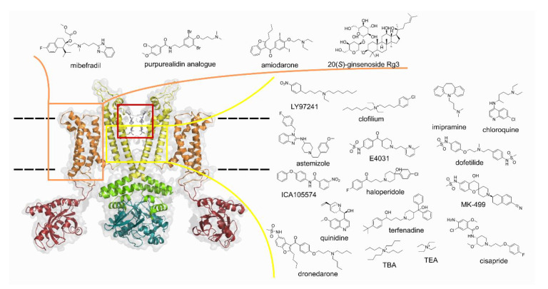
Figure 1.
Opposite two subunits of KV10.1 with voltage-sensor domain (VSD) in orange, pore domain and central cavity in yellow, selectivity filter in red box. Structures of known ligands binding to the VSD and the central cavity of the pore domain. The structure (PDB: 5k7l) in the figure represents Kv10.1 with the pore domain in the closed conformation due to calmodulin binding and VSD in the upward/conductive conformation. TBA: tetrabutylammonium: TEA: tetraethylammonium. The structure of KV10.1 was prepared using PyMOL [21].

