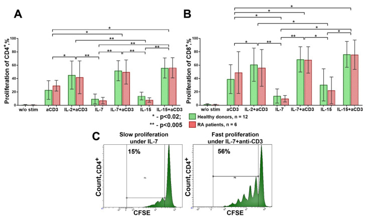
Figure 6.
Proliferation of CD4+ (A) and CD8+ (B) cells in healthy donors (n = 12) and RA patients (n = 6); (C) example of slow and fast proliferation (for an example of one of the donors). Significantly higher proliferation of CD4+ and CD8+ cells was observed when the influence of cytokines (IL-2, IL-7, or IL-15) was accompanied by TCR stimulation with anti-CD3 antibodies. Mean ± SD. A comparison of related groups was performed using one-way analysis of variance for dependent groups (RM one-way ANOVA), and post hoc analysis was performed using Tukey’s tests. Unrelated groups were compared using unpaired Student’s t-tests. RA, rheumatoid arthritis.

