Figure 4.
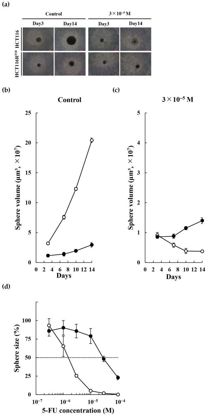
Tumor sphere formation of 5-FU-resistant HCT116RF10 and parental HCT116 cells. (a) Tumor sphere formation was analyzed using a Leica DMi1 microscope with 50× magnification. Scale bar = 500 μm. HCT116RF10 and parental HCT116 cells were treated with or without 3 × 10⁻⁵ M 5-FU for 3 or 14 days. Control, no 5-FU, solvent (DMSO) alone. To assess the ability of HCT116RF10 and parental HCT116 cells to form tumor spheres, the cells were treated with solvent alone (b) or 3 × 10⁻⁵ M 5-FU (c) for 14 days. Tumor sphere size was calculated as described in the Materials and Methods. White circle, HCT116 cells; black circle, HCT116RF10 cells. (d) Drug sensitivity of 5-FU in HCT116 and HCT116RF10 tumor spheres. Tumor sphere formation by HCT116RF10 and parental HCT116 cells after a 14-day treatment with 5-FU at the indicated concentrations. Results are the averages for groups of three tumor spheres each with error bars showing SE. White circle, HCT116 cells; black circle, HCT116RF10 cells.

