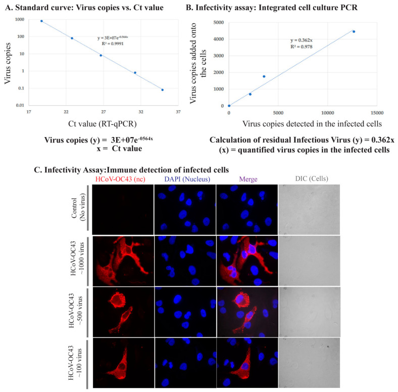
Figure 2.
Assay establishment: Integrated cell culture PCR and Immune detection of HCoV-OC43 infected cells. (A). Standard curve of varying HCoV-OC43 genome copies with Ct value. (B). Correlation of HCoV-OC43 viral copies added onto A549-hACE2 cells and detection of intracellular viral genome copies following infection and replication. (C). Immune localization of HCoV-OC43 nucleocapsid protein in A549-hACE2 cells infected with varying amounts of virus. Nucleocapsid was detected with anti-HCoV-OC43 antibody followed by staining with chicken anti-mouse Alexa Fluor 594 (red). Nuclei were stained with DAPI (blue). DIC images are to show cell morphology.

