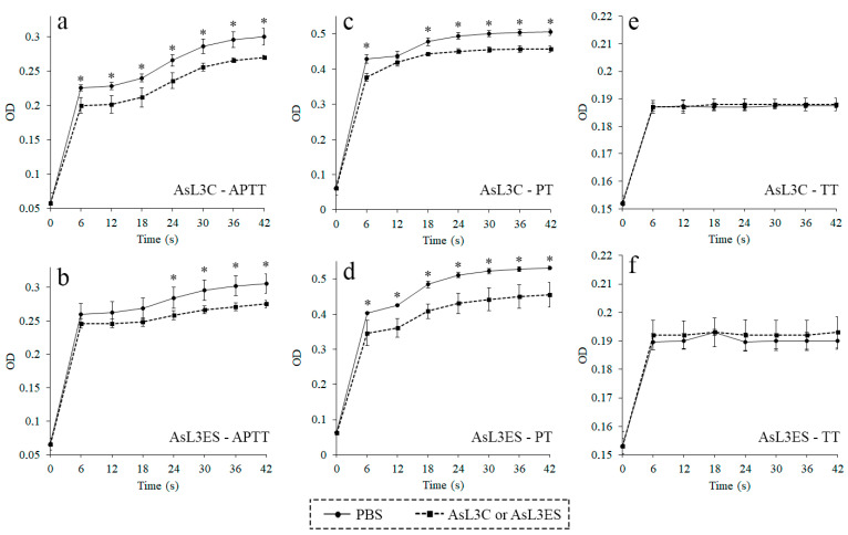
Figure 1.
Anticoagulant activity of AsL3C (a,c,e) and AsL3ES (b,d,f) evaluated by measuring the APTT (a,b), the PT (c,d) and the TT (e,f). Plasma from pigs was incubated with 0.5 μg of the antigenic extract (■) or with PBS as a negative control (●), and the corresponding reagent (APTT, PT or TT). Each point represents the mean of three replicates ± SD. Significant differences (p < 0.05) are marked with an asterisk (*).

