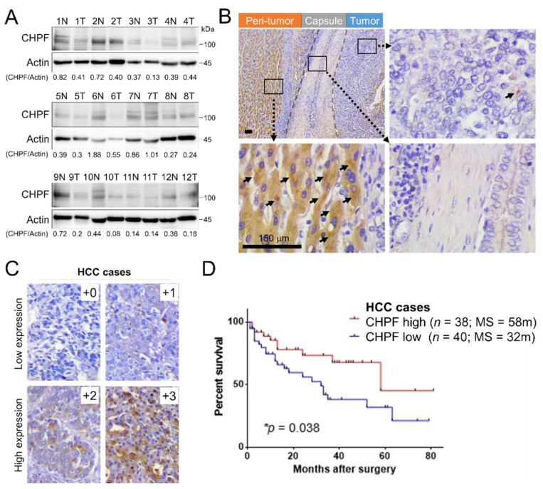
Figure 1.
CHPF expression in human hepatocellular carcinoma (HCC). (A) CHPF levels and quantification in non-tumor (peri-tumor) liver (N) and tumor tissue (T) of primary HCC tissues from twelve patients. (B) Immunohistochemistry of CHPF in a representative HCC tissue. Dot-like precipitations of CHPF (arrow) were observed mainly in peri-tumor tissues but rarely in stromal and tumor tissues. The scale bar indicates 50 μm. (C) Representative images of four grades of CHPF expression in primary HCC tissue (+0, negative; +1, <20%; +2, 20–50%; +3, >50%); n = 78. The scale bar indicates 150 μm. (D) Kaplan–Meier analysis of overall survival of HCC patients. The analyses were conducted according to the immunostaining of CHPF low expression (+0 and +1) and high expression (+2 and +3); p = 0.038.

