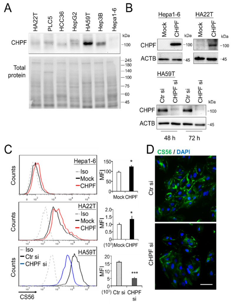
Figure 2.
CHPF expression in HCC cells. (A) CHPF expression in seven HCC cell lines. Protein expression was analyzed by Western blotting, and total protein was used as an internal loading control. (B) Stable CHPF overexpression in Hepa1-6 and HA22T cells. Transient CHPF knockdown in HA59T cells 48 and 72 h after siRNA transfection. Protein expression was analyzed by Western blotting, and β-actin (ACTB) was used as an internal control. (C) Surface CS56 antibody staining (an anti-chondroitin sulfate antibody) on Hepa1-6, HA22T, and HA59T transfectants was analyzed by flow cytometry with anti-mouse IgM-FITC. Nonspecific mouse IgM was used as an isotype control (Iso). * p < 0.05, *** p < 0.001. (D) Fluorescence microscopy analysis of CS56 (green); DAPI (blue) indicated the position of the nucleus. The scale bar indicates 50 μm. The cells were fixed with paraformaldehyde without permeabilization.

