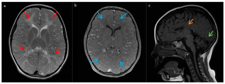
Figure 1.
MRI findings in the patient. These images, obtained at the age of 7, show: (a) Hypomyelination process with diffuse hyperintensity of the supratentorial and cerebellar white matter on the T2-weighted images (red arrows); (b) T1 diffuse isointensity of the supratentorial white matter on the T1 sequences (blue arrows); (c) Cerebellar atrophy (green arrow), and a thinned corpus callosum (orange arrow).

