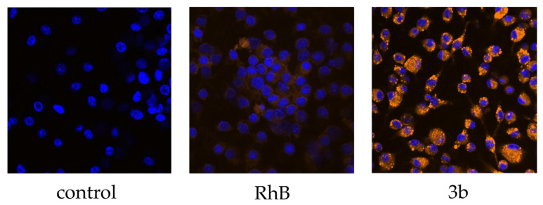
Figure 3.
Confocal microscope imaging of mouse bone marrow-derived macrophages untreated (control), treated with solution of free Rhodamine B in concentration 15 µg/mL (RhB) and with formulation 3b representing nanospheres fabricated from branched PLGA using NPM. Cells were incubated with the samples for 1 h. The nuclei were stained by dye Hoechst 33342. Magnification 40× was used.

