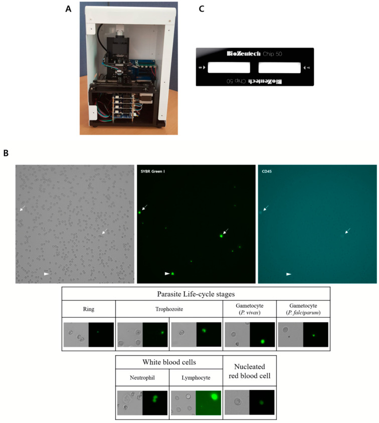
Figure 1.
The automated microscopic malaria parasite detection system. (A) Automated microscopic malaria parasite detection system hardware. (B) Image of stained malarial parasite-infected blood cells showing malarial parasites, white blood cells (WBCs) and nucleated red blood cells obtained using the automated microscopic malaria parasite detection system at 40× magnification (1200 × 1200 pixel2). The image is obtained at green LED (upper left), blue LED (upper middle) and red LED (upper right) are shown. The arrows indicate WBCs and the arrow heads indicate gametocyte. The example images of P. vivax at different stages, P. falciparum gametocyte and WBCs are also shown. (C) The plastic chip for the automated microscopic malaria parasite detection system.

