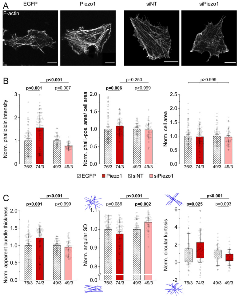
Figure 5.
Piezo1-dependent alterations of HAF stiffness are a result of differential organization of the actin cytoskeleton. (A) representative images of the actin cytoskeleton of HAF 3 days post-transfection, stained by phalloidin. Scale bars = 20 µm. (B) average phalloidin intensity, phalloidin-positive area per cell area, and cell area. (C) apparent thickness, angular standard deviation (SD), and circular kurtosis of actin bundles. All data normalized to the mean of the respective control. See Figure S3 for raw data. n/N = number of cells/number of experiments.

