Figure 3.
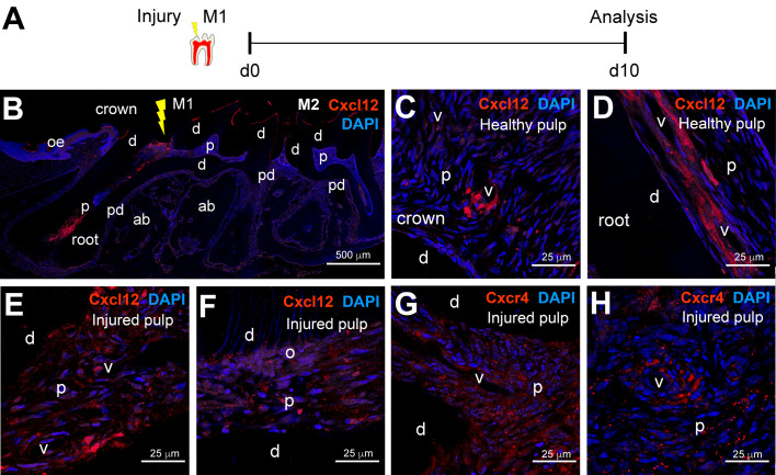
Distribution of Cxcl12 and Cxcr4 molecules in injured dental pulps. (A) Experimental design. (B) Immunofluorescent staining showing an overview of Cxcl12 protein distribution (red color) upon injury in molars. DAPI in blue color. The yellow arrow marks the injury site. (C,D) Immunofluorescent staining in dental pulp of intact healthy teeth showing Cxcl12 distribution (red color) in the crown (C) and the root region (D). DAPI in blue color. (E,F) Immunofluorescent staining showing Cxcl12 distribution (red color) in injured dental pulp; (E) shows a region of the injured pulp distant from the injury site; (F) shows Cxcl12 distribution in immediate proximity to the injury. (G,H) Immunofluorescent staining showing Cxcr4 distribution in injured dental pulp. Abbreviations: ab, alveolar bone; d, dentin; M1, first molar; M2, second molar; o, odontoblasts; oe, oral epithelium; p, dental pulp; pd, periodontium; v, vessels. Scale bars: (B), 500 μm; (C–H), 25 μm.

