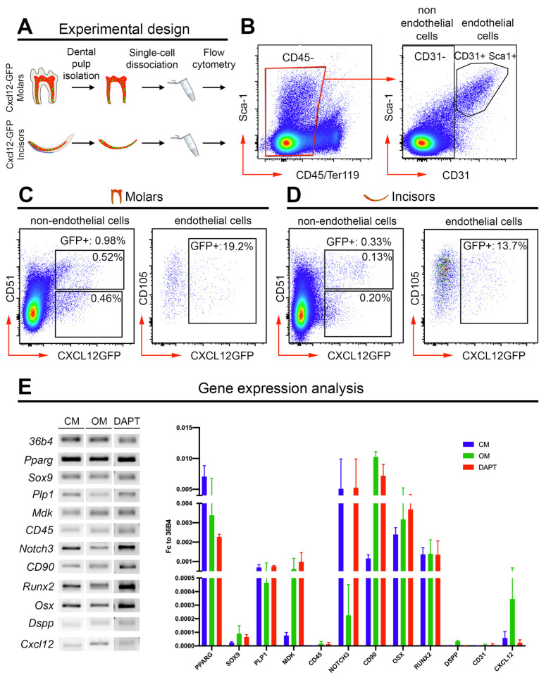
Figure 4.
Isolation and characterization of Cxcl12-GFP dental pulp cells. (A) Experimental design. (B) Isolation of nonimmune (CD45-) dental pulp cells and sorting of endothelial (CD31+) vs. nonendothelial (CD31-) dental pulp cells. (C) Fluorescence Activated Cell-Sorter (FACS) quantification of endothelial and nonendothelial Cxcl12-GFP+ cells in molar dental pulps. (D) FACS quantification of endothelial and nonendothelial Cxcl12-GFP+ cells in incisor dental pulps. (E) Gene expression analysis of nonendothelial (CD31-) Cxcl12-GFP+ dental pulp cells cultured in control medium (CM), osteogenic medium (OM), and in the presence of the Notch pathway inhibitor DAPT (DAPT). Gene expression data obtained from three independent biological replicates.

