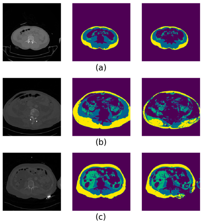
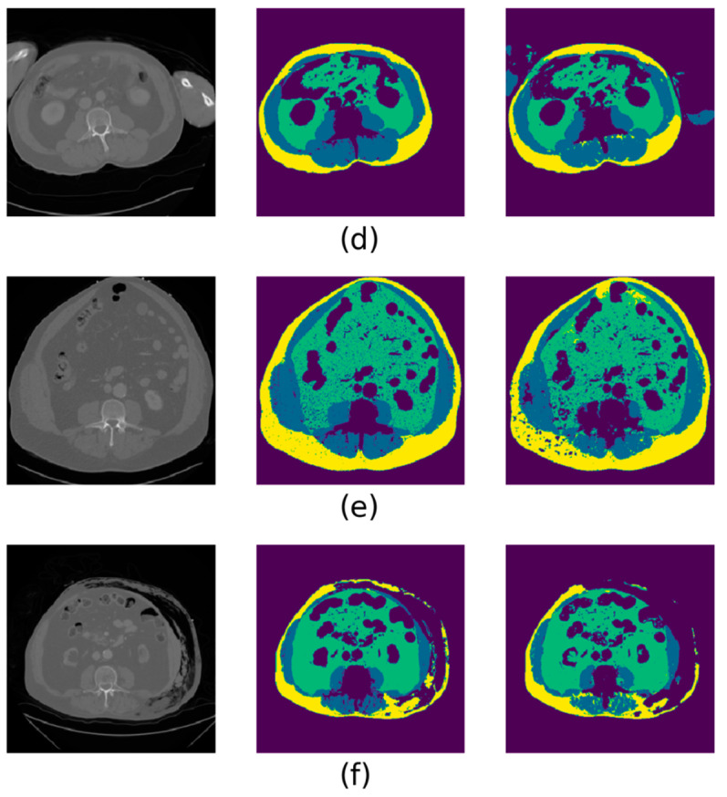
Figure 3.
Selected examples of deep-learning automated segmentation results, with representative errors represented. In each image, the original L3 CT slice is shown on the left, the ground truth segmentation in the middle and the automated segmentation on the right. The color scheme is as follows—yellow: subcutaneous adipose tissue; blue: lumbar muscle; green: visceral adipose tissue. (a) An automatic segmentation that would be deemed clinically acceptable. (b) A CT “streak” scatter artefact near the spine that led to internal organs and adipose being mislabeled. (c) A case of an unknown foreign object lying under the left dorsolateral side of the patient, creating strong scatter artefacts that led to the misclassification of subcutaneous adipose as muscle. (d) A common event in the trauma dataset that was only rarely seen in the training dataset, i.e., hands and arms in the CT field of view being misclassified as lumbar muscle. (e) A noisier CT image than usual, resulting in spots of undetected adipose and muscle. (f) A rare case of post-traumatic subcutaneous emphysema, leading to missed detection of subcutaneous adipose.


