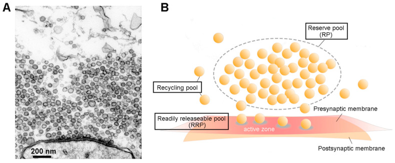
Figure 1.
Synaptic vesicles (SVs) and their pools. (A). Electron micrograph of an active zone (AZ) from a squid giant synapse. SVs are the dark, membrane-bound circles that are both attached to the presynaptic plasma membrane and clustered nearby. (B). Classification of SV pools. A reserve pool (RP) occupies the distal volume of the presynaptic terminal, while a readily releasable pool (RRP) consists of vesicles docked at the AZ. The other freely moving SVs are assigned to a recycling pool.

