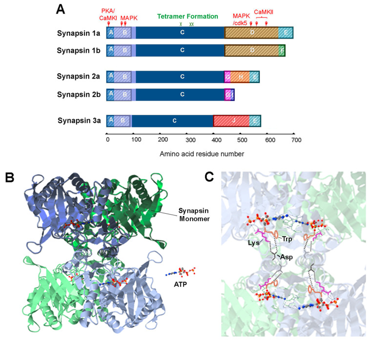
Figure 2.
Synapsin domain structure and tetrameric structure of synapsins. (A). Synapsin isoforms consist of numerous domains, indicated by colors and letters. IDR regions are indicated by shading. Adapted from [41]. (B). Tetrameric structure formed by the C domain of synapsin 2 in the presence of ATP. Two synapsin monomers in a dimer—colored blue or green—are shaded with dark or light colors. A tetramer consists of two dimers. (C). The key residues mediating tetramer formation: aspartate (Asp, black), tryptophan (Trp, orange) and lysine (Lys, magenta); bonds between synapsin dimers are indicated by dashed lines. Structures in (B,C) adapted from data in [42], (accessible at PDB ID: 1i7l), visualized with the NGL viewer [43].

