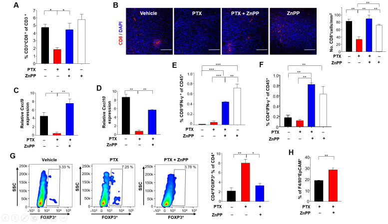
Figure 6.
Effects of HO-1 inhibition on T cell-mediated anti-tumor immunity in 4T1-tumor bearing mice under PTX therapy. (A) CD45+CD3+CD8+ tumor-infiltrating lymphocyte populations (as percentages) were identified by flow cytometry. (B) Representative images of tumor sections stained for CD8+ T cells and DAPI are shown. Scale bar, 200 μm. (C,D) The whole-tumor mRNA expression levels of Cxcl9 (C) and Cxcl10 (D) were analyzed by qPCR. (E) The percentages of IFN-γ+CD8+ T cells in CD45+ populations were detected by flow cytometry. (F) The proportions of IFN-γ+CD4+ T cells in the CD45+ populations were analyzed by flow cytometry. (G) The percentages of Foxp3+CD4+ T cells were analyzed by flow cytometry. (H) Engulfment of breast tumor cell debris was analyzed as the proportion of F4/80+ macrophages found to contain intracellular EpCAM (a standard epithelial tumor cell marker), as assessed by flow cytometry. *, **, *** Significantly different between groups compared (* p < 0.05; ** p < 0.01; *** p < 0.001).

