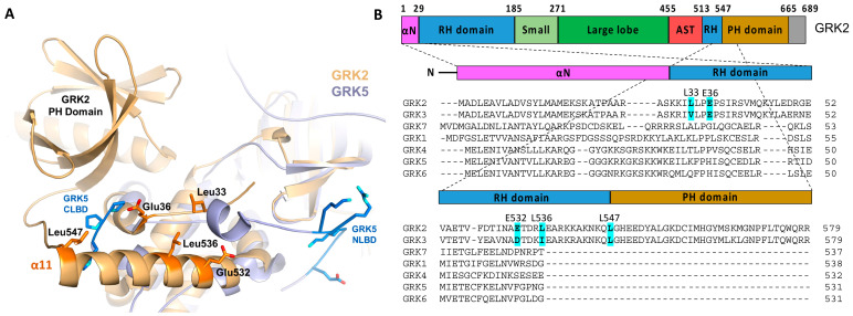
Figure 3.
Hypothetical GPCR-interacting sites in the GRK2 RH domain. (A) GRK2 (PDB: 4PNK [72], gold) aligned with full-length GRK5 (PDB: 4TND [73], slate) indicated the presence of a patch of solvent-exposed residues uniquely conserved in GRK2/3 and similarly disposed with respect to the membrane as the NLBD and CLBD of GRK5. These residues could conceivably interact with activated GPCRs [49]. Side chains of the GRK2 residues that were modified in this study are depicted as orange sticks. (B) Domain diagram of GRK2 and the structure-based sequence alignment of the seven human GRKs, with the targeted positions in GRK2 highlighted.

