Figure 4.
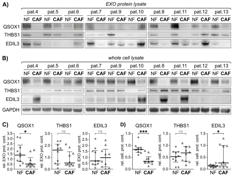
Exosomal fibroblast activity marker validation in an independent validation cohort. Twenty additional fibroblast cell lines derived from 10 CRC patients were subjected to cellular protein and EXO isolation. Exosomal protein was isolated and subjected to immunoblot. (A) Immunoblot analysis of quiescin sulfhydryl oxidase 1 (QSOX1), thrombospondin 1 (THBS1), EGF-like repeats and discoidin domains 3 (EDIL3), in primary fibroblasts-derived EXOs. (B) Immunoblot analysis of QSOX1, THBS1 and EDIL3 in primary fibroblasts whole cell lysate, including GAPDH as loading control. (C) Graphical analysis of EXO protein immunoblots shown in (A) using ImageJ, relative to GAPDH. Mann-Whitney-U test: * p < 0.05. (D) Graphical analysis of cellular protein immunoblots shown in (B) using ImageJ, relative to GAPDH. Mann-Whitney-U test: * p < 0.05, *** p < 0.001. ns = not significant.

