Figure 1.
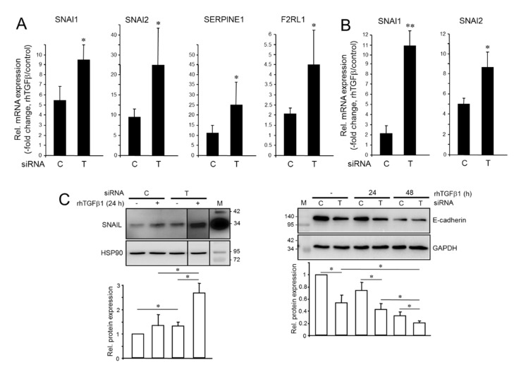
Effect of endogenous and recombinant human transforming growth factor β1 (rhTGFβ1) on gene expression in tumor cells with aTGFβ production. (A) Panc1TGFB1-KD cells were transiently transfected twice on 2 consecutive days with 50 nM each of siRNA directed against TGFB1 (T) or a scrambled control (C) siRNA and incubated for another 48 h. Cells were then treated with rhTGFβ1 for 24 h and analyzed by qPCR for expression of the indicated genes and TATA box-binding protein (TBP) as a reference gene. Data are displayed as fold induction by rhTGFβ1 treatment over non-treated controls (means ± SD; n = 3 (SNAI1, SERPINE1), n = 4 (SNAI2, F2RL1)). (B) As in (A) except that MDA-MB-231TGFB1-KD were analyzed. (C) Western blot analysis of SNAIL (left-hand blot) and E-cadherin (right-hand blot) in Panc1TGFB1-KD cells treated, or not, with rhTGFβ1. Detection of heat shock protein 90 (HSP90) or glyceraldehyde 3-phosphate dehydrogenase (GAPDH) served as a loading control. The graphs below the blots show data quantification from densitometric readings of band intensities (mean ± SD, n = 3). The asterisks denote significance. The vertical lines between lanes 3, 4, and 5 of the left blot indicate that irrelevant lanes have been removed. Successful knockdown of TGFB1 was verified by ELISA of secreted TGFβ1 in culture supernatants (Figure S2). M, molecular weight marker. Numbers to the right or left of the blots denote band sizes in kDa. *, p < 0.05; **, p < 0.01.

