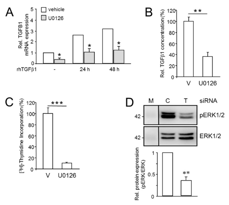
Figure 5.
Mutual regulatory interactions between aTGFβ1 and ERK signaling enhances proliferation. (A) Panc1 cells were treated, or not (-), for 24 or 48 h with rhTGFβ1 (5 ng/mL) in the absence or presence of U0126 (10 µM) or vehicle (0.1% dimethyl sulfoxide, DMSO) followed by qPCR analysis of TGFB1 and GAPDH and TBP as internal control. Data represent the normalized mean ± SD of triplicate wells from a representative experiment. The asterisks indicate significance relative to the vehicle ctrl. (B) As in (A), except that cells were switched to serum-reduced medium (0.5% FBS) prior to U0126 treatment. Cells were allowed to condition their growth media for 24 h. Aliquots of conditioned media were subjected to ELISA measurement of total (bioactive + latent) TGFβ1. Data are the mean ± SD of triplicate samples. (C) Panc1 cells were treated, for 24 h with U0126 (25 µM) or vehicle (V) followed by (3H)-thymidine incorporation. Data shown are the mean ± SD from 6 wells processed in parallel and are representative of 3 experiments. The asterisks indicate significance. (D) Panc1 cells were transfected with control or TGFB1 siRNA as described in the legend to Figure 1, 24 h later stimulated with rhTGFβ1 for 1 h and subjected to immunoblotting for phospho-ERK1/2 (pERK1/2), and total ERK1/2 as a loading control. Data represent the mean ± SD from three independent assays. Successful knockdown of TGFB1 was verified by ELISA of secreted TGFβ1. The asterisks (∗) indicate significant differences relative to the respective controls. *, p < 0.05; **, p < 0.01; ***, p < 0.001.

